
Mattek to Attend the 2026 Oral Health Research Congress
Mattek scientists will be attending and presenting posters at the Oral Health Research Congress in Lisbon, Portugal. Our scientists have 2 presentations on new research, applications, and oral tissue models. Visit them at Booth #10 to discuss your upcoming project needs!
Schedule a meeting with our teamPresentation:
Human Oral Tissue Model Detects Irritation Potential of Medical Devices
Marek Puskar1, Jennifer Molignano2, Christian Pellevoisin2, Silvia Letasiova1, Mitchell Klausner2
1. MatTek In Vitro Life Science Laboratories, Bratislava, Slovakia.
2. MatTek Corporation, Ashland, MA, United States.
Session: Oral Session – Oral Session 16 – Dental Materials: Biocompatibility, Bioengineering and Biologic Effects I
Location: Room 5 – Fernando Pessoa Room (Centro Cultural de Belém)
Time: September 3, 2026 | 4 pm to 6 pm
Presenter: Marek Puskar
Abstract
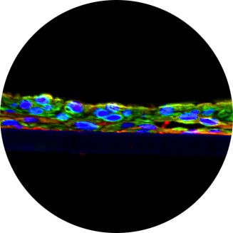
Objectives: Safety of any medical device (MD) designed for use in oral cavity has to be evaluated in accordance with ISO 10993-23. This standard specifies methods to assess oral mucosa irritation potential of MDs, by using in vivo Syrian hamster cheek pouch model. In contrast, our study focuses on development of alternative in vitro test method for assessment of oral mucosal irritation utilizing in vitro three-dimensional reconstructed tissue model of oral cavity, EpiOral. This model consists of normal, human-derived oral keratinocytes cultured to form stratified, highly differentiated model of the human buccal epithelium.
Methods: Preliminary tests were conducted to assess the feasibility of this in vitro approach. Chemicals commonly used in manufacture of MDs were dissolved at various concentrations in polar (saline) and nonpolar (sesame oil) solvents and applied topically to EpiOral tissues model for 1,4 and 18 hours. Irritation potential was evaluated by measuring the time required for a test substance to reduce tissue viability to 50% (ET-50) utilizing MTT colorimetric viability assay. To further enhance the robustness of the method, a single 18h exposure time point was selected for assessment of irritation and additional colorimetric viability assay utilizing WST-1 was included into testing.
Results: The results showed relationship between exposure time and viability of the cells as well as ET-50 and concentration of irritant chemical. Furthermore, the irritation potential of the tested substances at relevant concentrations was in accordance with historical in vivo data from Syrian hamster.
Conclusions: The data demonstrated that this in vitro assay performs equivalently to the in vivo method.
Poster Presentation:
In Vitro Wound Healing Models of Oral Mucosa (P330)
Emily, Reis1, Marek Puskar1, Jennifer Molignano2, Silvia Letasiova1, Alexander Armento2, Seyoum Ayehunie2, Mitchell Klausner2
1. MatTek In Vitro Life Science Laboratories, Bratislava, Slovakia.
2. MatTek Corporation, Ashland, MA, United States.
Session: September 5, 2026 | Presentation from 8:30am to 10:00 am
Presenter: Emily Reis
Abstract
Objectives: The epithelial lining of the oral cavity frequently suffers from wounds as result of surgeries, accidents or ulcers. A weakened epithelial barrier poses a risk of infection and facilitates the penetration of irritants, which can lead to delayed or impaired healing, increased pain, and potential spread of pathogens if not managed promptly. To address this, we developed an in vitro oral mucosal wound-healing models utilizing EpiOral and EpiGingival tissues. Both EpiOral and EpiGingival consist of normal, human-derived oral epithelial cells cultured to form multilayered, highly differentiated models of human buccal (EpiOral) and gingival (EpiGingival) phenotypes.
Methods: Wounds of 3mm diameter were incised into EpiOral and EpiGingival tissue models with circular biopsy punch. Wound re-epithelialization over time was evaluated by histological cross-sections, transepithelial electrical resistance (TEER), and bright-field (BF) microscopy. The toxicological properties of the tissues were assessed by determining the ET-50 following exposure to 1% sodium dodecyl sulfate (SDS).
Results: Histological analyses and BF microscopy showed that wound closure occurs within 2 days in EpiOral and within 4 days in EpiGingival tissues. Results of TEER measurements supported these findings. After an initial decrease in TEER values post-wounding a progressive increase in TEER was observed over time course. Wounding also altered the toxicological profile of the tissues. Following exposure to 1% SDS, the ET-50 values of wounded EpiOral tissue model decreased by 43.4% compared with non-wounded tissues. In the cornified EpiGingival model,a 56.2% reduction of ET-50 values was measured.
Conclusions: In conclusion, these findings demonstrate the successful development of wounded oral mucosa tissue models. These models can serve as valuable tool for evaluating new therapeutic compounds aimed at accelerating oral wound closure and for determining toxicity profiles in barrier-compromised oral mucosa.