
Drug Metabolism
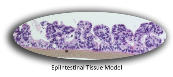
EpiIntestinal is a highly differentiated 3D tissue model that closely recapitulates the physiology, tissue structure, and function of the epithelium of the small intestine. Produced from normal human cells, EpiIntestinal is used in pharmaceutical development applications including the prediction of drug absorption and metabolism.
The Model
The EpiIntestinal tissue model is produced at the air-liquid-interface (ALI) in easy-to-handle tissue culture inserts and 96-well HTS screening plates. Characterization of the EpiIntestinal tissue model demonstrates the presence of columnar shaped, proliferative basal cells (including LGR5+ Adult Stem Cells), Paneth Cells, M Cells and Enterocytes along with the expression and activity of efflux transporters. Structural analysis of EpiIntestinal confirms a polarized epithelium with villi structures and Kerckring folds. Ultrastructurally, EpiIntestinal exhibits brush borders, functional tight junctions and mucous secreting granules, similar to in vivo tissue.
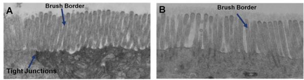
Ultrastructural Analysis: Transmission electron micrograph (TEM) of A) in vitro EpiIntestinal and B) Explant tissue showing Brush borders (situated at the luminal pole of the enterocyte) and tight junctions. Brush border provides digestive and absorption surface; site for enzymes & transporters.
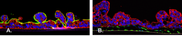
Characterization: Cross-sectional microscopic images of A) EpiIntestinal (epithelium only) labeled for CK19 (Red, epithelial cell marker) and Villin (Green, brush border marker) and B) EpiIntestinalFT (full thickness tissue containing fibroblasts) labeled for Vimentin (Green, fibroblast marker) and CK19 (Red).
EpiIntestinal Drug Metabolism
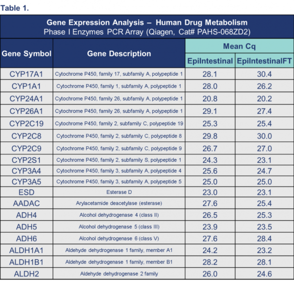
For orally administered drugs, the small intestine can be a major site for presystemic drug metabolism. Initial studies show that the EpiIntestinal microphysiological tissue model expresses levels of CYP3A4 comparable to the Jejunum region of the human small intestine (Figure 1). Gene expression analysis of EpiIntestinal also shows the expression of many other phase I enzymes important for drug metabolism (Table 1).
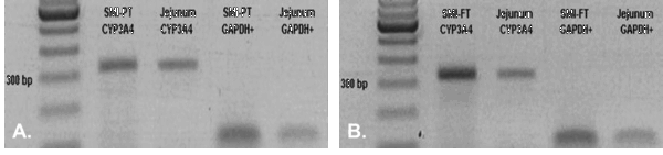
Figure 1: Gene expression analysis showing the expression of Cytochrome P (CYP) 3A4 expression in A) EpiIntestinal (epithelium only) and B) EpiIntestinalFT (full thickness tissue containing fibroblasts) compared to human small intestine explant tissue (Jejunum).
For more information, view the EpiIntestinal Technology Page, and Technical References.
Request a Quote
Thank you for requesting information about Mattek products! A representative will contact you shortly.
**If you would like to place an order for Mattek products, please contact Customer Service**