
Multiplexed Immunofluorescent Custom Panel Services
Biomarker profiling of tissues with 10+ markers on a single slide using our multiplex immunolabeling and imaging services
A deeper understanding of cellular interactions within the context of cancer and inflammatory diseases is critical to developing better treatments and therapeutic outcomes. With advances in fluorescent probes, imaging technologies and image analysis, fluorescent immunohistochemistry is a powerful tool to probe cell populations and cell interactions within tissues. Visikol’s multiplexed immunofluorescent and imaging services allows for biomarker profiling of tissues with up to 15 markers on a single slide.
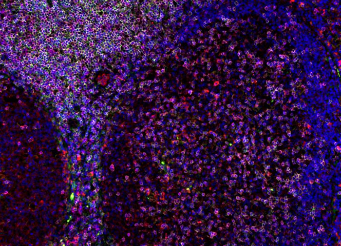
Immunofluorescence staining in FFPE human tonsil of CD3 (magenta), CD4 (green), and PDL1 (red).
Workflow
- Send Your Tissues
- Send us your fixed tissue samples, FFPE tissue blocks, or unstained slides.
- Choose Your Panels
- Select the markers for your specific research needs. You can choose up to 15 markers.
- Panels will be comprised of 3-4 antibodies.
- QC of client tissue
- Validation of antibodies
- 3 concentrations will be tested for each antibody, and a secondary only control is included as well.
- The best concentration will be chosen and shared with the client prior to moving to the next step.
- Multiplex Fluorescent Immunohistochemistry
- Label samples with each panel (a nuclear counterstain included in each panel).
- Slides can be imaged at 10x, 20x, or 40x magnification.
- After each round of labeling the previous panel of antibodies will be stripped using Visikol’s proprietary EasyPlex™ technology.
- Co-registration of panels
- Each panel image for a sample is co-registered using custom-built image processing algorithms to create a multiplexed fluorescence image stack.
- A single image with every marker will be uploaded to Visikol’s cloud sharing platform, BitSlide™ for each sample.
- Analytical endpoints
- Various analytical endpoints can be chosen based on the research question.
- Cell population quantification
- Area analysis
- Spatial analysis
Rapid, inexpensive, high-plex microenvironment and biomarker profiling of tissue with any antibody.
Cutting-Edge Tools
Using our proprietary EasyPlex™ antibody stripping reagent, multiple rounds of immunolabeling can be conducted on a single slide, providing unprecedented detail about your samples.
High Value Endpoints
Using digital slide imaging and advanced image analysis, a large number of valuable endpoints can be captured from a single specimen, giving unique insights about cell populations and interactions.
Take Action
Our expert team processes images and analyzes data, delivering actionable insights in an easy-to-read report, so you can make decisions and push forward your research efforts.