
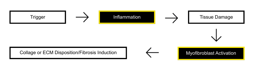
Intestinal Fibrosis
The EpiIntestinal full thickness (SMI-FT) tissue model is useful for testing candidate drugs or biologics to treat diseases that are characterized by inflammation of the small intestine.
The Model
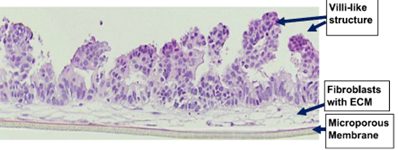
EpiIntestinal Full Thickness (SMI-FT) is a 3D reconstructed tissue model produced from normal, human cell-derived small intestine epithelial cells and fibroblasts. The highly differentiated tissue model is produced at the air-liquid-interface (ALI) in easy-to-handle tissue culture inserts. Structural analysis of the tissue model demonstrates columnar shaped basal cells and Kerckring folds. Ultrastructurally, EpiIntestinal exhibits brush borders, functional tight junctions and mucous secreting granules, similar to in vivo tissue.
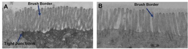
Transmission electron micrograph (TEM) of in vitro EpiIntestinal (A) and Explant tissues (B) showing Brush borders (situated at the luminal pole of the enterocyte) and tight junctions. Brush border – provides digestive and absorption surface; site for enzymes & transporters.
The Method
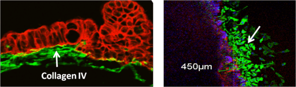
SMI-100-FT Tissue Model SMI-100-FT Tissue Model – Wounded
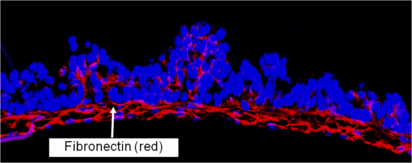
SMI-100-FT Tissue model stained for ECM protein – Fibronectin (red) and DAPI nuclear stain (blue).
For more information, view the EpiIntestinal and References.
Request a Quote
Thank you for requesting information about Mattek products! A representative will contact you shortly.
**If you would like to place an order for Mattek products, please contact Customer Service**
Questions?
sales@mattek.com
+1-508-881-6771