Cell Viability
Visikol provides cytotoxicity assay services. Assessment of cell viability in 3D models is an excellent way to evaluate the cytotoxic effect of drug compounds.
Background
- The Ki-67 protein is a cellular marker for proliferation (1). During interphase, the Ki-67 antigen can be exclusively detected within the cell nucleus, whereas in mitosis most of the protein is relocated to the surface of the chromosomes (2). Ki-67 protein is present during all active phases of the cell cycle (G1, S, G2, and mitosis), but is absent in resting (quiescent) cells (G0) (3). Cellular content of Ki-67 protein markedly increases during cell progression through S phase of the cell cycle (4). Ki67 index is frequently cited by pathologists when diagnosing degree and severity of cancer patients to identify those who derive greater benefit from chemotherapy (5).
- Ki-67 is an excellent marker to determine the growth fraction of a given cell population. The fraction of Ki-67-positive tumor cells is often correlated with the clinical course of cancer. The prognostic value of Ki67 for survival and tumor recurrence have repeatedly been proven.
- In the screening of antiproliferative drugs for cancer, Ki67 is a commonly used marker to determine the fraction of cells currently in a proliferative state. Antiproliferative drugs such as cisplatin and paclitaxel markedly decrease the fraction of proliferating cells as measured by Ki67.
- 3D cell culture models more closely recapitulate the in vivo tumor environment, and are an effective tool for the evaluation of antiproliferative effects of promising anti-cancer drug compounds.
- Using High Content Screening with 3D cell models offers a medium-throughput approach to screening the antiproliferative activity of test compounds.
General Procedure
- For 3D cell models, grown to approximately 200 μm in diameter. Adherent monolayers cultured to confluence.
- Treatment with test compounds
- After 24 hours, cells are labeled with fixable dead cell probe.
- Nuclei are labeled with DAPI
- For 3D cell models, tissue clearing is applied to render 3D cell models transparent
- High content imaging is conducted on well plates
- Images are analyzed to quantify total number of cells and total number of dead cells
Protocol
| Instrument | Molecular Devices ImageExpress Micro Confocal |
| Analysis Method | High content screening |
| Markers | LIVE/DEAD fixable dead cell probe (Molecular Probes) DAPI (total cell count) Other dead cell probes available on request |
| Cell Model Type | 3D cell models (e.g. tumor spheroids, organoids, etc.) or adherent monolayer / cell suspension |
| Cell Types Available | HepG2, HepaRG, primary human hepatocytes ATCC cancer cell lines (e.g. A549, BT549, MCF-7) Primary cells Client provided cells iPSC derived neural organoid (Stemonix) Custom models available on request |
| Test Article Concentration | 8 point assay (0.05, 0.1, 0.5, 1, 5, 10, 50, 100 µM) (custom concentrations available) Single point assay |
| Number of Replicates | 3 replicates per concentration |
| Quality Controls | 0.5% DMSO (vehicle control) Staurosporin (positive control) |
| Test Article Requirements | 50 uL of 20 mM solution or equivalent amount of solid |
| Data Delivery | Dose response curves, LD50 values, total cell counts, Viability % for each test concentration Evaluation of statistical significance of results with respect to vehicle and positive control |
| Assay Reference Code | INV-2102 |
Validation Data

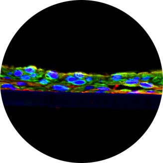
Figure 1. Maximum Z-projection of Z-stack obtained from High Content Imaging of HepG2 tumor spheroids treated with cisplatin. Data was quantified by automated image analysis to count total number of cells, and number of dead cells

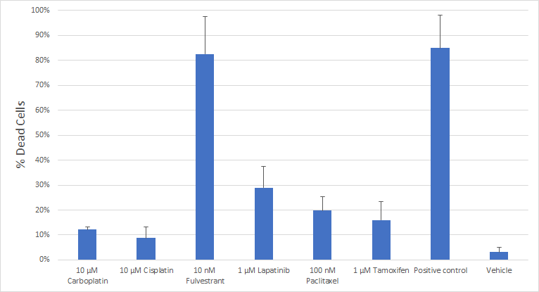
Figure 2. Representative data showing the observed fraction of dead cells detected in primary tumor spheroids treated with various cytotoxic agents
References
- Wiederschain, G.Y. Biochemistry Moscow (2011) 76: 1276. https://doi.org/10.1134/S0006297911110101

