
Immunofluorescent Pre-Validated Panel Services
Pre-validated panels that simplify the immunofluorescent labeling process.
How it Works
Pre-validated panels simplify the immunofluorescent labeling process by providing ready-to-use, optimized marker combinations that streamline workflows and accelerate biological insights. Visikol’s multiplexed immunofluorescent and imaging services allows for biomarker profiling of tissues with up to 15 markers on a single slide.
Workflow
- Send Your Tissues
- Send us your fixed tissue samples, FFPE tissue blocks, or unstained slides.
- Choose Your Panels
- Select from the pre-validated human panels.
- Looking for non-human species? Check out our custom IF services.
- Can select multiple panels if desired.
- Select from the pre-validated human panels.
- QC of client tissue
- Multiplex/Fluorescent Immunohistochemistry
- Label samples with each panel (a nuclear counterstain included in each panel).
- Slides can be imaged at 10x, 20x, or 40x magnification.
- For multiple panels, antibodies will be stripped using Visikol’s proprietary EasyPlex™ technology after each round of labeling
- Co-registration of panels (if applicable)
- All panel images for a sample are co-registered using custom-built image processing algorithms to create a multiplexed fluorescence image stack.
- A single image with every marker will be uploaded to Visikol’s cloud sharing platform, BitSlide™, for each sample.
- Analytical endpoints
- Various analytical endpoints can be chosen based on the research question.
- Cell population quantification
- Area analysis
- Spatial analysis
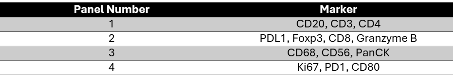
Figured 1. Immunofluorescent staining in FFPE human tonsil of Panel 1. CD3 (yellow), CD4 (red), CD20 (purple)
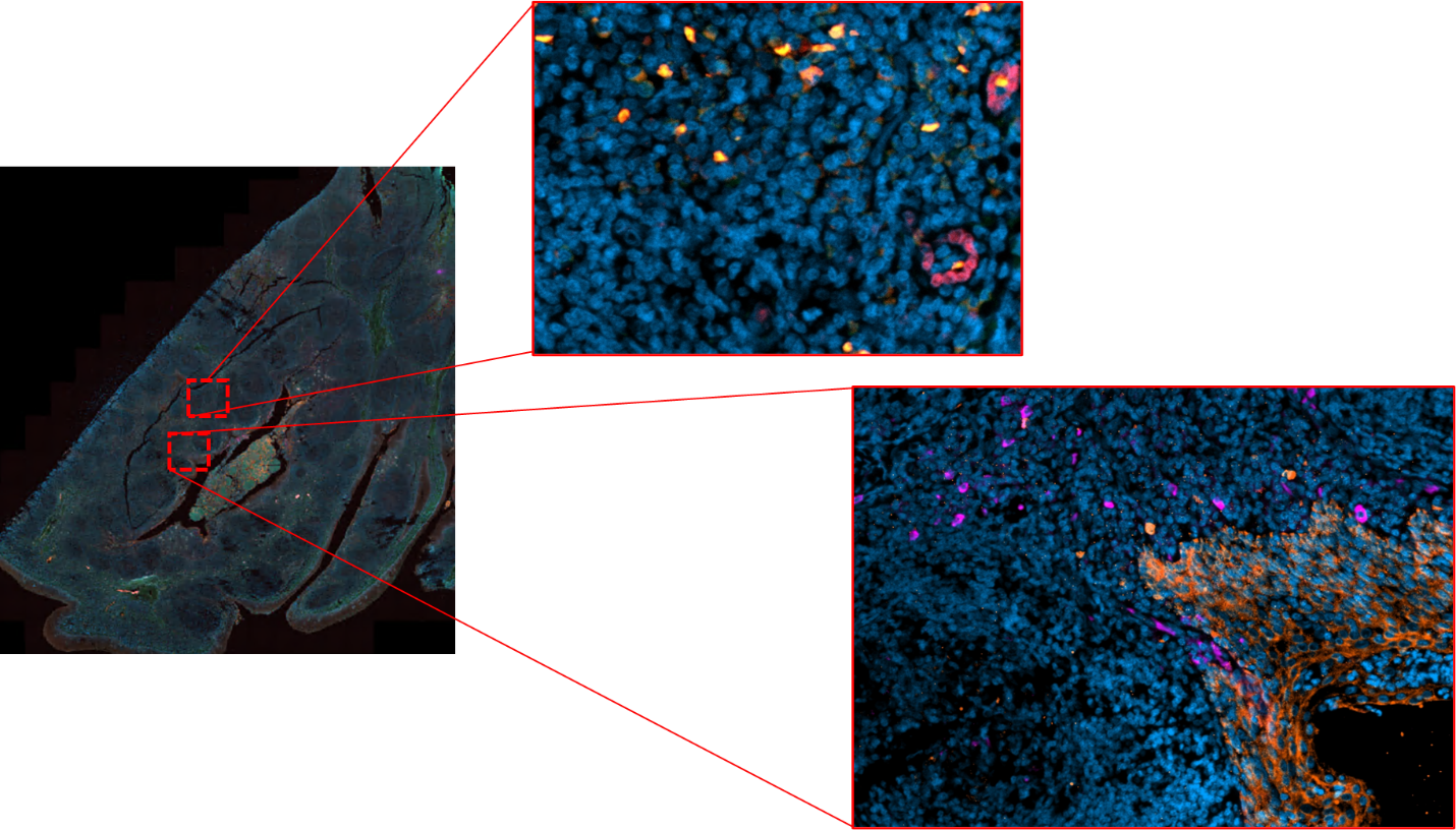
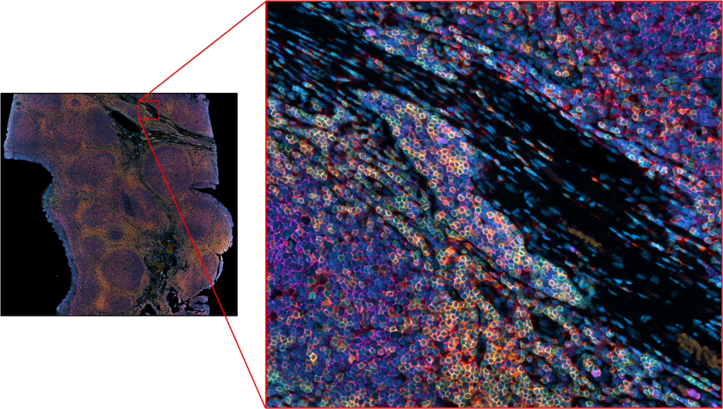
Figure 2. Immunofluorescent staining in FFPE human tonsil of Panel 2. PDL1 (purple), Granzyme B (red), FoxP3 (green); Yellow represents the co-expression of Granzyme B and FoxP3.
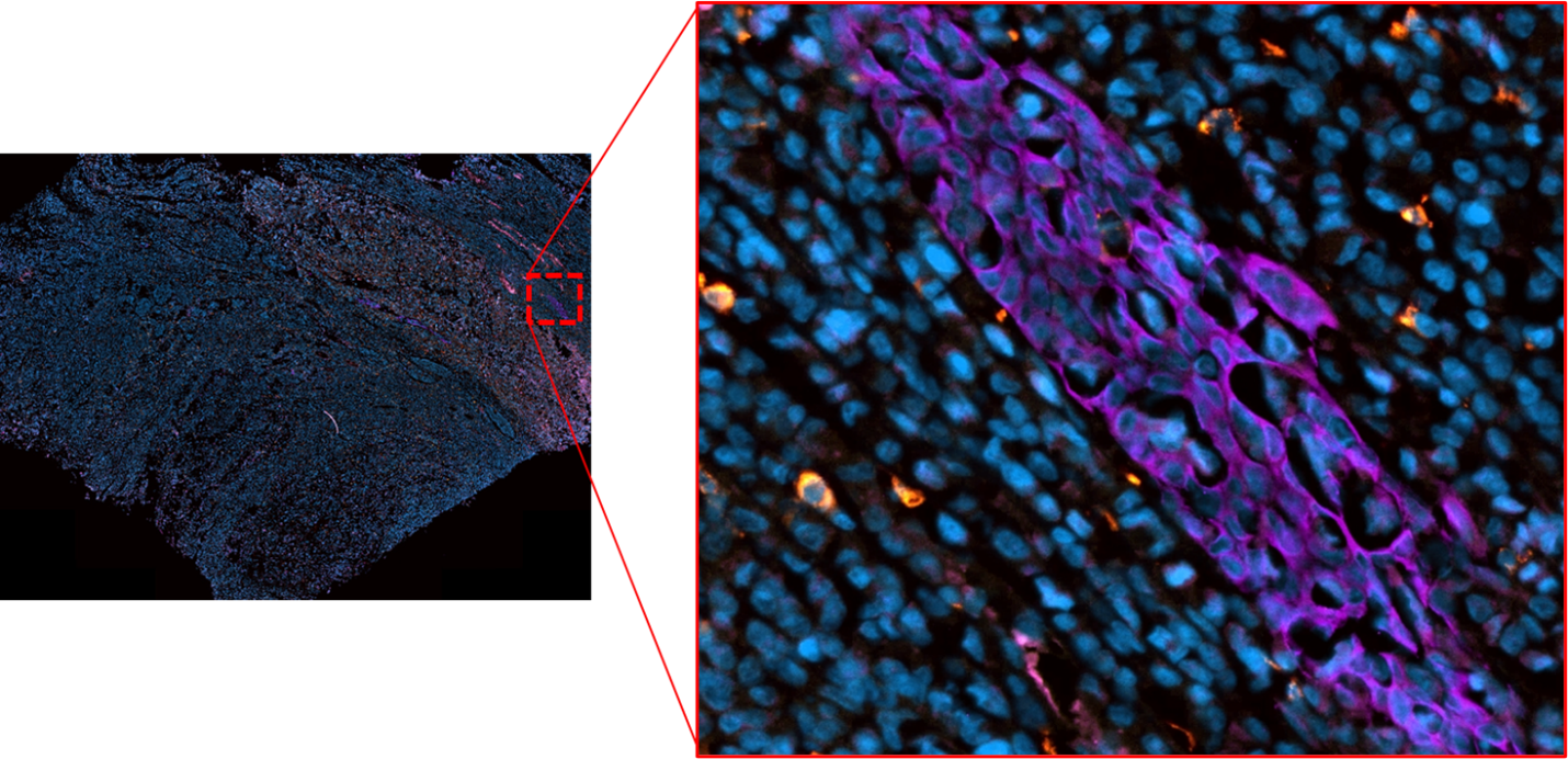 Figure 3. Immunofluorescent staining in FFPE human tonsil of Panel 3. CD56 (red), CD68 (yellow), PanCK (purple); Orange represents the co-expression of CD56 and CD68.
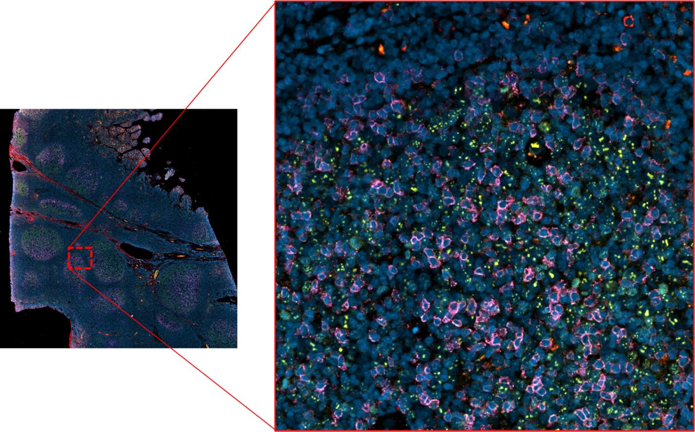 Figure 4. Immunofluorescent staining in FFPE human tonsil of Panel 4. Ki67 (green), PD1 (red), CD80 (purple)